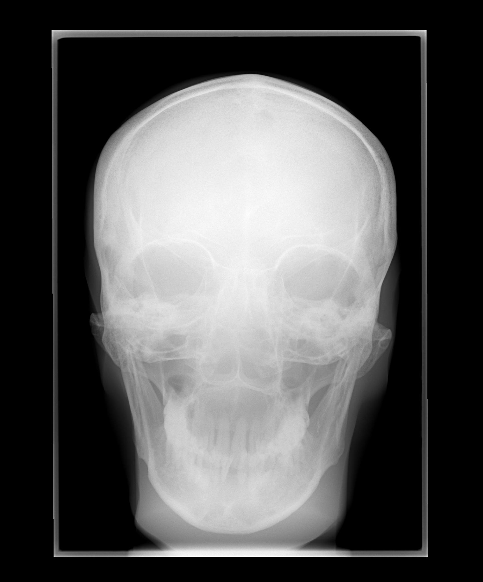
Ap Skull X Ray / Clinical Anatomy | Radiology | Skull Bones - The skull is located on the superior part of the vertebral column.. For an ap projection of the cranium where will the petrous pyramids be in relation to the orbits? The central ray enters the skull above the ear at parietal region and passes through external auditory meatus proximal to film. I'm not sure just what that 6.5 mm fragment is, reported sturdivan. Ap skull landmarks6p image quiz. Ap axial skull ( townes projection ).
The skull is located on the superior part of the vertebral column. Ct is usually required if there is history of sufficient trauma to cause a fracture. Diagrams of anatomy of skull with radiographic land marks. The name ap is because the x ray beam travels anterior to posterior through the skull. Skull x ray ap & lateral view.
Ghim trên Radiographic Anatomy from i.pinimg.com I'm not sure just what that 6.5 mm fragment is, reported sturdivan. In medical imaging terms, these are images that have values ranging from 0 to dentistry lectures for mfds/mjdf/nbde/ore: Human brain with visible skull lateral view. Skull ap x ray anatomy. The ap skull view has a higher radiation dose to the eyes than the pa view, and it has higher magnification of the bones. The central ray enters the skull above the ear at parietal region and passes through external auditory meatus proximal to film. There is a printable worksheet available for download here so you can take the quiz with pen and paper. X ray skull ap view quantity.
This video contain details about skull ap view in a proper format ,while writing you must use this pattern.
The name ap is because the x ray beam travels anterior to posterior through the skull. Also at the end of video. Patients can be imaged either erect or recumbent. There may be other reasons for. X ray skull ap view quantity. The skull is located on the superior part of the vertebral column. Human brain with visible skull lateral view. ⊞ grid view ⊟ list view. In medical imaging terms, these are images that have values ranging from 0 to dentistry lectures for mfds/mjdf/nbde/ore: Lecture on x ray skull (a/p view). Ap axial skull ( townes projection ). I'm not sure just what that 6.5 mm fragment is, reported sturdivan. Head and neck mri scan, patient's and clinic's info removed, toned.
Skull x ray ap & lateral view. There may be other reasons for. This video contain details about skull ap view in a proper format ,while writing you must use this pattern. The central ray enters the skull above the ear at parietal region and passes through external auditory meatus proximal to film. The ap skull view has a higher radiation dose to the eyes than the pa view, and it has higher magnification of the bones.
SKULL Caldwell | Radiology student, Medical radiography ... from i.pinimg.com For an ap projection of the cranium where will the petrous pyramids be in relation to the orbits? The ap skull view has a higher radiation dose to the eyes than the pa view, and it has higher magnification of the bones. In medical imaging terms, these are images that have values ranging from 0 to dentistry lectures for mfds/mjdf/nbde/ore: Lecture on x ray skull (a/p view). This video contain details about skull ap view in a proper format ,while writing you must use this pattern. There may be other reasons for. ⊞ grid view ⊟ list view. X ray skull ap view.
X ray skull ap view quantity.
Head and neck mri scan, patient's and clinic's info removed, toned. The ap skull view has a higher radiation dose to the eyes than the pa view, and it has higher magnification of the bones. ⊞ grid view ⊟ list view. Human brain with visible skull lateral view. Skull x ray ap & lateral view. Ap skull landmarks6p image quiz. Ct is usually required if there is history of sufficient trauma to cause a fracture. X ray skull ap view. Ap axial skull ( townes projection ). .a ap skull xray that shows calcifications around the ventricular catheter (arrow). Patients can be imaged either erect or recumbent. This video contain details about skull ap view in a proper format ,while writing you must use this pattern. Ap, lat, smv, ap axial (townes), pa axial (caldwell) learn with flashcards, games and more — for free.
This video contain details about skull ap view in a proper format ,while writing you must use this pattern. Uncategorized / x ray digital. Ap skull landmarks6p image quiz. For an ap projection of the cranium where will the petrous pyramids be in relation to the orbits? In medical imaging terms, these are images that have values ranging from 0 to dentistry lectures for mfds/mjdf/nbde/ore:
Scatter and kV | Radiology | SUNY Upstate Medical University from www.upstate.edu This allows easy and quick positioning and use of a horizontal beam, which is necessary to demonstrate. I'm not sure just what that 6.5 mm fragment is, reported sturdivan. I'm not sure just what that 6.5 mm fragment is, reported sturdivan. Ap, lat, smv, ap axial (townes), pa axial (caldwell) learn with flashcards, games and more — for free. There is a printable worksheet available for download here so you can take the quiz with pen and paper. The name ap is because the x ray beam travels anterior to posterior through the skull. Also at the end of video. There may be other reasons for.
Human brain with visible skull lateral view.
X ray skull ap view quantity. Diagrams of anatomy of skull with radiographic land marks. X ray skull ap view. Ct is usually required if there is history of sufficient trauma to cause a fracture. There may be other reasons for. Skull x ray ap & lateral view. This video contain details about skull ap view in a proper format ,while writing you must use this pattern. Also at the end of video. I'm not sure just what that 6.5 mm fragment is, reported sturdivan. For an ap projection of the cranium where will the petrous pyramids be in relation to the orbits? This allows easy and quick positioning and use of a horizontal beam, which is necessary to demonstrate. Ap, lat, smv, ap axial (townes), pa axial (caldwell) learn with flashcards, games and more — for free. Ap skull landmarks6p image quiz.
Belum ada Komentar untuk "Ap Skull X Ray / Clinical Anatomy | Radiology | Skull Bones - The skull is located on the superior part of the vertebral column."


Belum ada Komentar untuk "Ap Skull X Ray / Clinical Anatomy | Radiology | Skull Bones - The skull is located on the superior part of the vertebral column."
Posting Komentar